An epithelium is a special arrangement of cells that line many parts of our body including the skin as well as lining any tubular structures in the body. Generally speaking epithelium makes up the 'functional' parts of all of our organs. An epithelium rests on a basement membrane and is defined as an avascular tissue, meaning no vessels run through it. The cells that make up epithelial tissue are situated closely together as sheets or glands that separate a surface from underlying tissue. Because of their function, epithelial cells have a few prime distinguishing features. On this page you'll read more about:
- Characteristic features of epithelium
- How to categorise epithelial tissue
- Specialisations of the apical domain
- Cell to cell adhesions
- Glands
CHARACTERISTIC FEATURES OF EPITHELIUM
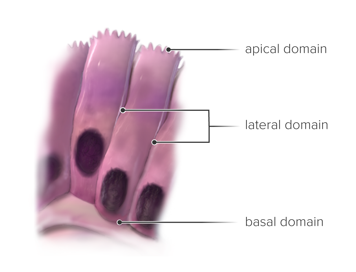
Firstly, epithelial cells are carefully placed side to side through specific cell-to-cell adhesion molecules. These molecules form specialised cell junctions. Epithelium creates a selective barrier between the external environment and the underlying connective tissue. It facilitates or inhibits the passage of specific substances between the exterior environment and the underlying connective tissue compartment. The different domains of the epithelial cell and their names (basal, apical and lateral) are illustrated in the image on the right.
HOW TO CATEGORISE EPITHELIAL TISSUES
Terminology is based on structure. The classification of epithelium is based on two principles:
THE NUMBER OF CELL LAYERS
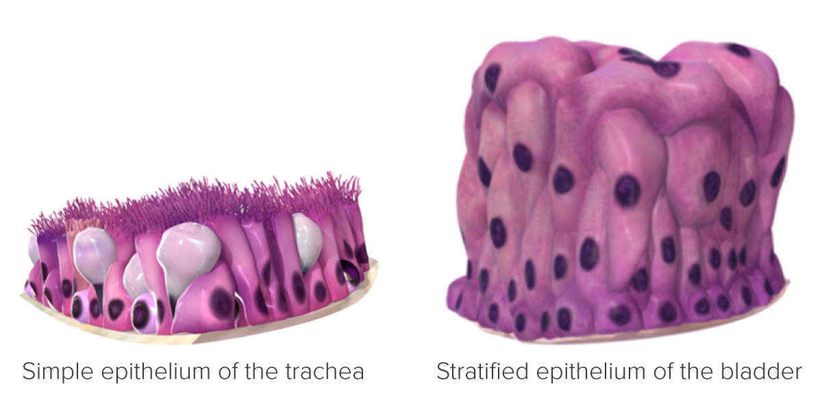
Simple epithelium is one cell layer thick. Stratified epithelium has two or more cell layers. Pseudostratified epithelium appears stratified but when viewed in three dimensions all of the cells, at some point, touch the basement membrane.
THE SHAPE OF THE SURFACE CELLS
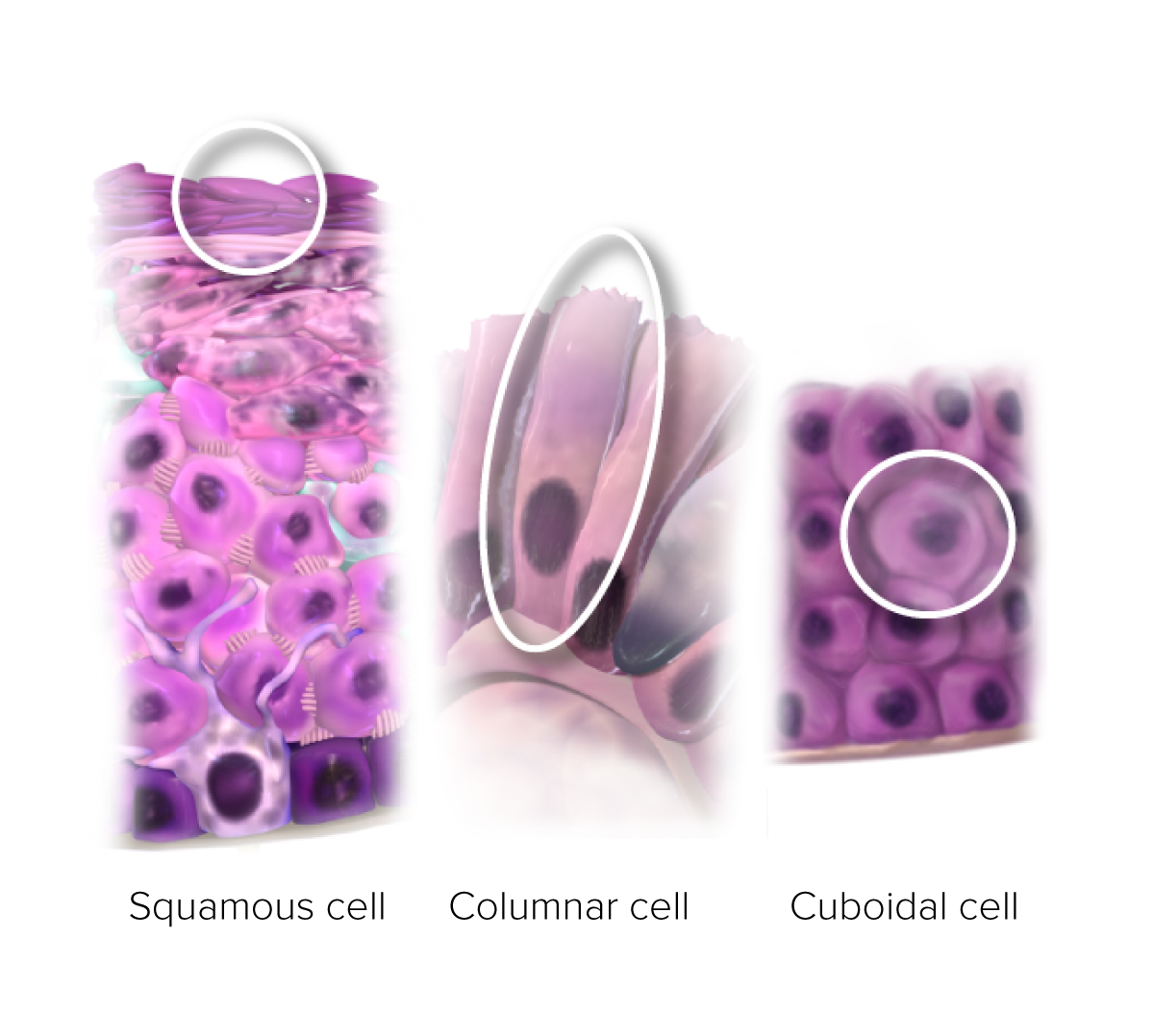
The individual cells that compose an epithelium are described as cuboidal: the width, depth and height are approximately the same; columnar: the height of the cell clearly exceeds the width; squamous: The width and length clearly exceeds the hight of the cell.
In a stratified epithelium, the shape and height of the cells usually varies from layer to layer, but only the shape of the cells that form the surface layer is used when classifying the epithelium.
Transitional epithelium (urothelium) is stratified and lines the lower part of the urinary tract. Cells on its free surface layer transition form large, round, dome-shaped cells to squamous cells depending on the distention of the organ.
Squamous epithelial cells are generally wider than they are high but squamous epithelium is morphologically defined by the presence of keratin production or intracellular prickles. These two morphological features are of great importance when making a diagnosis of squamous cell carcinoma.
SPECIALISATIONS OF THE APICAL DOMAIN
The apical domain of epithelial cells is situated at the top of the epithelium towards the external environment or luminal edge. Some apical cells show special features giving them important homeostatic functions.
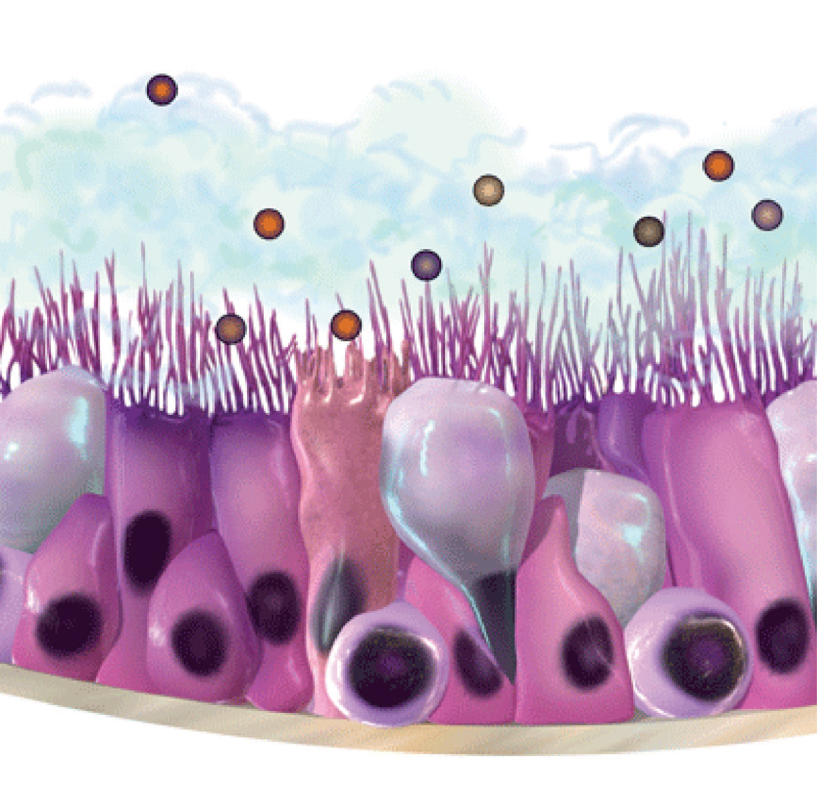
Microvilli: in order to increase the apical surface area for absorption, some epithelial cells have small, finger-like processes that have a core of actin filaments. under a light microscope, these microvilli can be seen as a 'brush border'
Motile cilia: These are hair-like extensions of the apical plasma membrane. Their function is the movement of particles. They can be found in tracheal epithelium.
CELL TO CELL ADHESIONS IN THE LATERAL DOMAIN
A characteristic feature of the lateral domain of epithelial cells is the presence of cell adhesion molecules that form junctional complexes between the apposed lateral domains of neighbouring cells. There are several types of junctions; namely occluding, anchoring or communicating junctions. These junctions can be viewed on the right.
GLANDS
There are two types of glands one can observe in the epithelium:
Exocrine glands secrete their products directly onto a surface.
Endocrine glands lack a duct system. They secrete their products (hormones) into the bloodstream to reach a specific receptor on distant target cells.
Exocrine glands can be classified as mucous glands (mucous secretions) and serous glands (protein-rich and watery secretions). There are three ways in which exocrine glands secrete products: merocrine (exocytosis), apocrine (product is released in vesicles containing a thin layer of cytoplasm), and holocrine (the product is accompanied by cell debris from the dying secretory cell). These mechanisms are illustrated on the left.
SEE FOR YOURSELF
Want to have a look at the world through a microscope? Log in to the virtual microscope. Use your University of Dundee login information and have a look at a collection of microscopy images of different kinds of epithelium.