INTERACTIVE 3D MODEL OF THE DUODENUM
For more information on how to navigate the model, click on the question mark icon on the bottom right corner of the viewing screen.
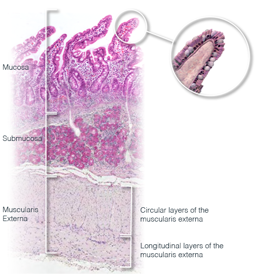
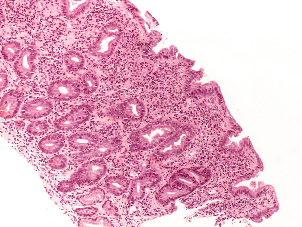
EPITHELIUM OF THE DUODENUM
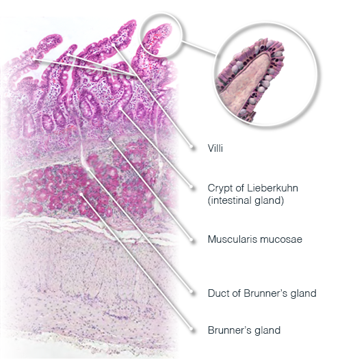
The duodenum, like the remainder of the small bowel, consists of tall villi projecting into the lumen of the small bowel. This greatly increases the surface area available and allows for more absorption than would otherwise be possible. The distinctive feature of the duodenum is the submucosal Brunner's glands. These glands produce an alkaline mucin which protects the bowel against leakage of acid from the stomach.
Also present within the duodenum are a variety of neuroendocrine cells that aid in the digestive process. Cholecystokinin (CCK) is secreted by such cells in response to food entering the duodenum. CCK has many functions including inhibition of gastric acid secretion as well as stimulating release of digestive enzymes from the pancreas.
Throughout it’s length the bowel is exposed to numerous external antigens and so associated with all parts of the bowel are a population of immune cells. Scattered lymphocytes can be seen within the epithelium itself and the lamina propria of the bowel contains a striking number of inflammatory cells even in normal conditions.
CLINICAL CONTEXT - MALABSORPTION
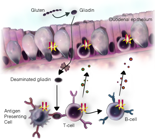
One of the commonest causes of malabsorption in the West is coeliac disease. Coeliac disease is caused by an abnormal immune reaction to gliadin in wheat. Because this is an abnormal immune reaction there is a dramatic increase in inflammatory cells (particularly T lymphocytes) within the lamina propria and epithelium itself. As part of this inflammatory reaction the epithelium is damaged and the villi become shorter and shorter and in severe cases absent. This “villous atrophy” can be seen on biopsies in this condition. Furthermore, the symptoms of diarrhoea are more easily understood when we appreciate that the surface area for absorption is reduced with loss of villi
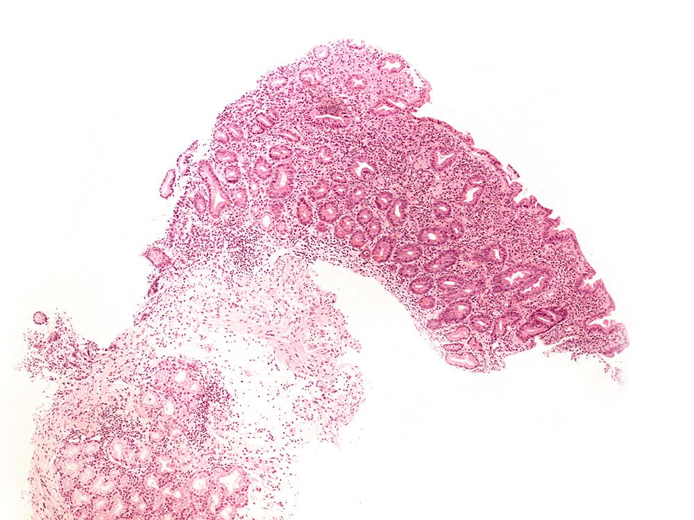
Below are two images of the duodenum in coeliac disease. The left hand image is at lower magnification and when compared to the normal images above it is easy to identify that the villi are not present. In fact, it is only the presence of the underlying Brunner's glands that allow you to confidently say this represents duodenum at all. This loss of epithelial surface area greatly reduces the absorptive capacity of the bowel and given this image it comes as no surprise that the symptoms and signs of coeliac disease are a result of diarrhoea and malabsorption.
The second image is at a higher magnification and shows that, along with loss of villi, there is a marked increase in intraepithelial inflammatory cells. In coeliac disease, the majority of these are T-lymphocytes.
SEE FOR YOURSELF
Want to have a look at the world through a microscope? Log in to the virtual microscope. Use your University of Dundee login information and have a look at a collection of microscopy images of different kinds of epithelium.
