INTERACTIVE 3D MODEL OF THE COLON
For more information on how to navigate the model, click on the question mark icon on the bottom right corner of the viewing screen.
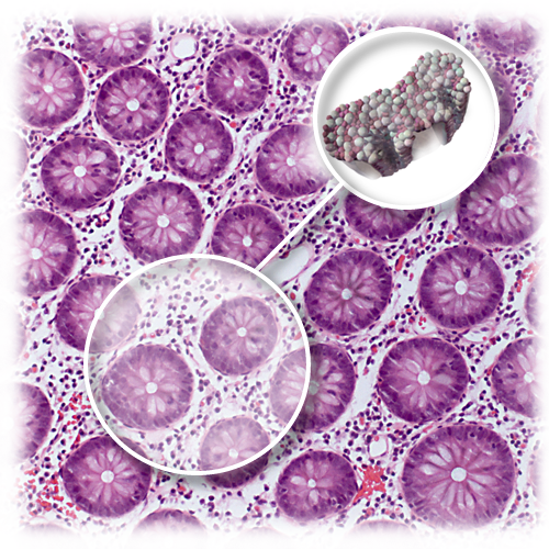
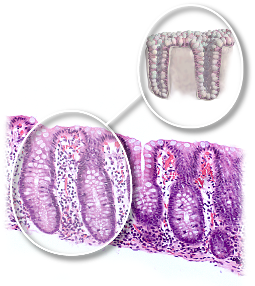
EPITHELIUM OF COLON
In contrast to the small bowel the epithelial surface of the large bowel is flat. Instead of having villi projecting from the surface, the large bowel has crypts that descend down from the surface throught the lamina propria to touch the base of the epithelium lying on top of a thin layer of muscle known as the muscularis mucosae. Colonic crypts contain a mixture of absorptive cells and a large number of goblet cells. Goblet cells produce large volumes of mucous which is released onto the surface of the epithelium creating a layer of lubrication to aid in the passage of feaces.
As with other areas of the bowel a striking number of immune cells are present within the large bowel to aid in processing of the many foreign antigens present within bowel contents. Read more about the surrounding structures of the epithelium of the colon or malignancies of the colon.
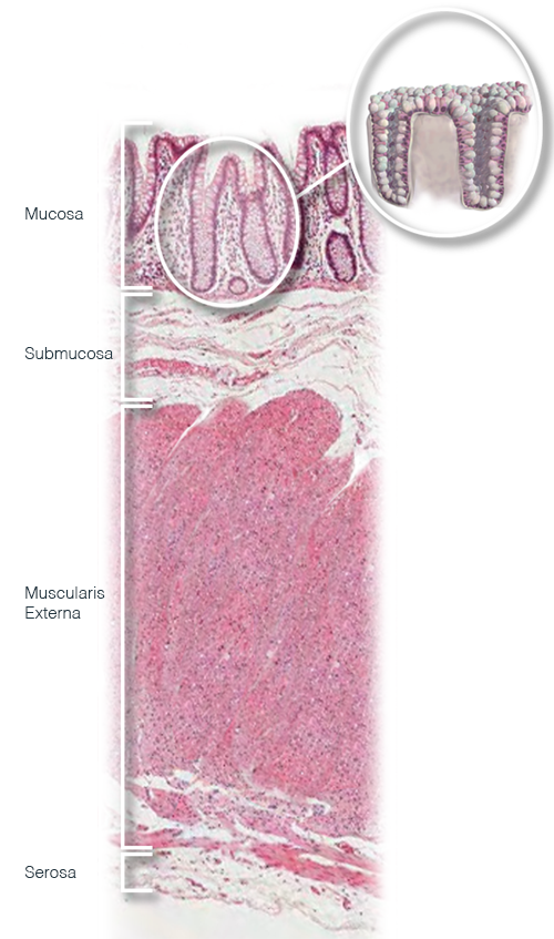
SURROUNDING STRUCTURES
In the images on the right and below microscope images of the structures surrounding the model can be viewed, as well as the situation of the epithelium in relation to them.
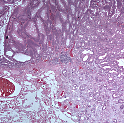
ADENOCARCINOMA VERSUS NON-INVASIVE TUMOR
Morphological identification of the layers of the bowel wall is important in distinguishing between pre-malignant and malignant lesions in the bowel. There are many types of polyp in the bowel, some of which show abnormality as a result of genetic mutation and are referred to as dysplastic. Dysplastic lesions are pre-malignant, in this context meaning non-invasive. Dysplastic glandular lesions are referred to as adenomas. After accumulating more genetic errors these adenomas gain the ability to invade tissue at which point they are malignant and are referred to as adenocarcinomas. In the large bowel we define invasion as abnormal cells moving beyond the basement membrane and muscularis mucosae at the base of the epithelium.
The image on the right shows evolution from premalignant dysplastic change (adenoma) to malignancy (adenocarcinoma). Note that the malignant part of the lesion has extended below the basement membrane and muscular mucosae. The adenoma on the left is well defined and you can clearly draw a line encircling the abnormal glands. In contrast the invasive component on the lower right is less organised and appears more chaotic.
SEE FOR YOURSELF
Want to have a look at the world through a microscope? Log in to the virtual microscope. Use your University of Dundee login information and have a look at a collection of microscopy images of different kinds of epithelium.